For decades, the search for liver cancer has relied heavily on a single protein in the blood. That protein — alpha-fetoprotein, or AFP — became the standard marker used to monitor patients at risk for hepatocellular carcinoma. Clinicians measured it, guidelines recommended it, and patients were told a normal result was reassuring.
The problem is that AFP misses liver cancer in nearly half the people who have it.
This is not a fringe finding. It is a well-documented limitation that has driven significant research investment into understanding what other biological signals the body produces when liver cancer is present — and whether those signals can be measured more reliably. The science of liver cancer tumor markers has advanced considerably beyond AFP. Here is what that research has produced, and what it means for patients at elevated HCC risk.
What Are Tumor Markers?
Tumor markers are biological substances — typically proteins, hormones, enzymes, or genetic material — that are produced by cancer cells or by the body in response to cancer. They are measured in blood, urine, or tissue samples and used to support clinical decision-making in several ways:
- Identifying patients who may need further diagnostic investigation
- Monitoring patients with known cancer for treatment response
- Detecting potential recurrence after treatment
- Assessing risk in high-risk patient populations
Importantly, no tumor marker currently used in clinical practice is diagnostic by itself. A tumor marker result is a signal — one piece of information that a physician interprets alongside imaging, clinical history, and other test results to form a complete picture. This distinction matters enormously when understanding what liver cancer tumor markers can and cannot tell you.
AFP as a Liver Cancer Tumor Marker
Alpha-fetoprotein (AFP) is a protein normally produced by the developing foetus. After birth, AFP levels in healthy adults drop to very low levels. When liver cancer develops, AFP production can increase significantly — which is why elevated AFP became a useful screening signal for hepatocellular carcinoma.
AFP testing for HCC has been in clinical use since the 1970s. It is inexpensive, widely available, and remains part of many standard surveillance protocols recommended for high-risk patients — particularly those with cirrhosis or chronic hepatitis B or C.
For the populations AFP works in, it provides real clinical value. The challenge is that those populations are smaller than many patients — and some clinicians — assume.
Why One Marker Is Not Enough
AFP has a well-documented accuracy problem. Studies consistently show that AFP detects liver cancer in approximately 50 to 60 percent of cases. More strikingly, between 15 and 30 percent of HCC patients have completely normal AFP values throughout the entire course of their disease — meaning AFP would never have flagged them at all, no matter how often it was measured.
Several factors explain why AFP falls short as a standalone liver cancer tumor marker:
Tumour heterogeneity. Not all HCC tumours produce AFP. Some subtypes — particularly well-differentiated early-stage tumours — produce little to no AFP even when they are actively growing.
False positives from other liver conditions. AFP can be elevated in cirrhosis, active hepatitis, and non-cancerous liver conditions — not just cancer. This means an elevated AFP result does not confirm HCC, and a normal AFP result does not rule it out.
Stage sensitivity. AFP performs better at detecting advanced HCC than early-stage disease. Since early detection is where survival benefit is greatest, AFP’s limitations are most damaging precisely where they matter most.
According to the American Association for the Study of Liver Diseases, AFP alone is no longer recommended as a standalone surveillance tool. Current guidelines recommend AFP combined with ultrasound — an acknowledgement that AFP needs supplementation.
Newer Biomarker Approaches in Liver Cancer Research
The limitations of AFP have driven active research into additional liver cancer tumor markers that may provide better sensitivity, specificity, or both. Several approaches have emerged from this research.
AFP-L3 is a specific subfraction of AFP that is more closely associated with HCC than total AFP. Studies suggest AFP-L3 may have higher specificity for liver cancer, though it still carries accuracy limitations similar to standard AFP.
DCP (des-gamma-carboxyprothrombin) — also called PIVKA-II — is a protein produced by liver cancer cells that is distinct from AFP. Some research suggests DCP may detect HCC in patients with normal AFP levels, making it a potentially complementary marker.
The GALAD score combines AFP, AFP-L3, DCP, age, and sex into a single risk score. Clinical trials are underway examining its performance compared to ultrasound-based surveillance, though it is not yet commercially available to consumers.
RNA fusion transcripts represent a fundamentally different approach to liver cancer tumor markers — one that does not measure proteins at all, but instead analyses abnormal RNA molecules produced when chromosomal rearrangements occur in cancer cells. These transcripts are cancer-specific in a way that protein markers like AFP are not, which is why this approach has shown significantly higher accuracy in early detection research.
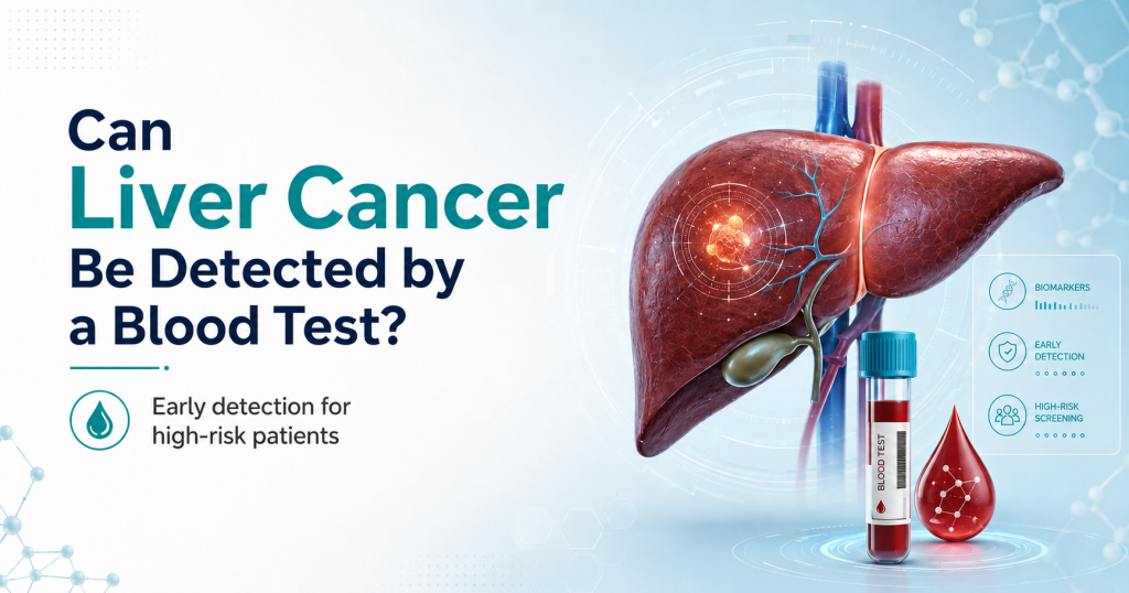
How MoleculeDx Uses Blood-Based Signals
MoleculeDx offers Fusion-detect — a blood-based HCC screening test built on NCI-funded research at the University of Pittsburgh School of Medicine — that moves beyond traditional liver cancer tumor markers by analysing RNA fusion transcripts directly.
The test identifies nine specific fusion transcripts — MAN2A1-FER, PTEN-NOLC1, SLC45A2-AMACR, and six others — that serve as highly specific molecular signals for hepatocellular carcinoma. Because these transcripts are produced by cancer-specific chromosomal events rather than general liver stress responses, they are not elevated by cirrhosis, hepatitis, or other benign liver conditions in the same way AFP is.
In NCI-supported research published in the American Journal of Pathology, Fusion-detect™ achieved up to 95 percent accuracy in detecting HCC — including in patients with completely normal AFP levels, the exact group that standard tumor marker testing fails most consistently.
About Fusion-detect
- 95% HCC detection accuracy
- Analyses RNA fusion transcripts — distinct from AFP
- Detects HCC in patients with normal AFP values
- Results within 24 hours
- At-home blood draw or walk-in at any UPMC location
- Available across all 50 US states
- $160 for at-home collection
Fusion-detect™ is a screening support tool. Results should be reviewed with a qualified healthcare provider as part of a broader HCC surveillance plan. The National Cancer Institute continues to fund research in this space as part of efforts to improve early liver cancer detection outcomes nationally.
Questions to Ask Your Healthcare Provider
If you are at elevated risk for HCC — due to cirrhosis, chronic hepatitis, NAFLD, or another liver condition — consider raising these questions at your next appointment:
- Which liver cancer tumor markers are appropriate for my specific risk profile?
- Is AFP alone sufficient for my surveillance, or should I be considering additional markers?
- Are there blood-based molecular tests that may add accuracy to my current plan?
- What does a normal AFP result actually mean given my underlying condition?
- How often should my markers be measured, and when would imaging be triggered?
These questions move the conversation from passive monitoring to active, informed surveillance — which is where early detection actually happens.
Frequently Asked Questions
What tumor marker is used for liver cancer? AFP (alpha-fetoprotein) is the most widely used liver cancer tumor marker in clinical practice. It has been part of HCC surveillance protocols since the 1970s and is typically measured alongside abdominal ultrasound every six months in high-risk patients. Newer approaches — including RNA fusion transcript analysis — are expanding what blood-based liver cancer detection can achieve beyond what AFP alone can offer.
Can AFP be high without cancer? Yes. AFP can be elevated in a range of non-cancerous conditions including liver cirrhosis, active hepatitis, non-alcoholic fatty liver disease, and other benign liver conditions. An elevated AFP result does not confirm a liver cancer diagnosis. Follow-up imaging — typically contrast-enhanced CT or MRI — is required to investigate an elevated AFP finding.
Can AFP be normal with liver cancer? Yes — and this is one of the most clinically significant limitations of AFP as a liver cancer tumor marker. Between 15 and 30 percent of HCC patients have completely normal AFP values throughout their disease. AFP-negative liver cancer is well-documented and is precisely why newer molecular approaches like Fusion-detect™ were developed — to detect HCC in patients that AFP would miss entirely.
Are tumor markers diagnostic by themselves? No. No liver cancer tumor marker currently in clinical use is sufficient to diagnose hepatocellular carcinoma on its own. Tumor markers are screening and surveillance tools — they identify patients who may need further investigation. A liver cancer diagnosis requires imaging such as contrast-enhanced CT or MRI, and in some cases biopsy. Tumor marker results should always be interpreted by a qualified physician in the context of the patient’s full clinical picture.
Explore Whether MoleculeDx Is Right for You
AFP has been the standard liver cancer tumor marker for fifty years. The science has moved forward. If you are at elevated risk for liver cancer and want to understand what blood-based screening options are now available beyond AFP, check your eligibility for Fusion-detect and book your test