Hepatocellular carcinoma (HCC) is the most common form of primary liver cancer. One of the major challenges in HCC is that early disease may cause few or no symptoms. Yet early detection matters because patients with localized disease have substantially more treatment options than patients whose cancer is discovered at an advanced stage.
People at increased risk for HCC—including many patients with cirrhosis, chronic hepatitis B or C, and other forms of chronic liver disease—may therefore undergo regular surveillance.
Ultrasound and the blood biomarker alpha-fetoprotein (AFP) have long played important roles in HCC surveillance. However, neither approach detects every early cancer. This has created considerable interest in new blood-based molecular markers that may provide information different from conventional protein biomarkers.
One emerging approach is the detection of cancer-associated fusion transcripts in blood.
What Is AFP, and Why Are Additional Blood Markers Needed?
AFP is a protein that can be measured in blood. AFP levels are elevated in some patients with HCC, but not in all of them. Published studies have reported substantial variation in AFP sensitivity and specificity depending on tumor size, patient population, and the cutoff used to define an abnormal result. The 2024 American Journal of Pathology study underlying part of the MoleculeDx research program noted reported AFP sensitivities of 49%–71% for HCC tumors smaller than 5 cm.
This does not mean that AFP has no value. Rather, it illustrates an important principle in cancer detection: a single protein marker may not capture the full molecular diversity of a cancer.
MoleculeDx research has therefore investigated a different type of signal—fusion transcripts released into the circulation.
What Are Fusion Genes and Fusion Transcripts?
Human genes normally occupy defined locations within chromosomes. In cancer cells, chromosomes can undergo rearrangements in which DNA segments that are normally separate become joined together.
Some of these rearrangements create a fusion gene.
When a fusion gene is transcribed into RNA, it produces a corresponding fusion transcript. In simple terms, a fusion transcript is an abnormal RNA molecule containing sequences derived from two genes that have become joined.
This distinction is important:
Fusion gene refers to the underlying DNA rearrangement.
Fusion transcript refers to the RNA produced from that rearranged gene.
Chromosomal rearrangements can generate recurrent fusion genes, and selected fusion transcripts can be detected as cell-free RNA in serum.
This creates an opportunity for a different kind of blood-based cancer marker: rather than measuring the concentration of a normal human protein such as AFP, researchers can look for abnormal molecular signals associated with cancer biology.
Can Fusion Transcripts Be Detected in the Blood of Patients With Liver Cancer?
Research from the University of Pittsburgh group that formed the scientific foundation for MoleculeDx has investigated this question for more than a decade.
A 2019 study published in Oncotarget1 examined fusion transcripts in serum from patients with HCC. The investigators demonstrated that selected cancer-associated fusion transcripts could be detected in serum and that serum-positive signals corresponded to fusion-positive HCC tissue in matched samples, supporting the tumor origin of these circulating RNA signals. The study also showed that individual fusion transcripts differed substantially in how readily they could be detected in blood.
That observation led to an important concept:
A multi-marker strategy may be more informative than relying on a single fusion transcript.
Different HCC tumors can contain different molecular alterations, and different fusion RNAs can also differ in their stability and detectability in circulation. The MoleculeDx research program therefore evolved toward measuring combinations of fusion-transcript signals.
From a Single Fusion Transcript to a Multi-Marker HCC Panel:
The current Liver Cancer Fusion Predictor analyzes nine cancer-associated fusion-gene transcripts in serum:
MAN2A1-FER, CCNH-C5orf30, SLC45A2-AMACR, PTEN-NOLC1, STAMBPL1-FAS, PCMTD1-SNTG1, ZNF124-SMYD3, VAPB-GNAS, and ZMPSTE24-ZMYM4.
These are not measured by next-generation sequencing in the clinical assay.
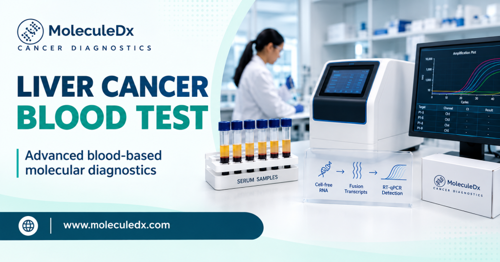
Instead, RNA is extracted from serum, converted to complementary DNA, and the fusion-junction sequences are detected and quantified using TaqMan real-time quantitative PCR.
This is an important distinction because the purpose of the test is not to sequence the patient’s genome. It is to measure a defined panel of specific RNA signals in a blood specimen.
Why Combine Multiple Molecular Signals?
Cancer is heterogeneous. Two patients with HCC do not necessarily have exactly the same molecular abnormalities.
For that reason, the MoleculeDx approach does not require every patient to have the same fusion transcript. Instead, quantitative information from multiple markers can be evaluated together.
A 2024 study published in The American Journal of Pathology2 evaluated serum fusion transcripts in HCC and non-HCC individuals and investigated logistic-regression machine-learning models for combining the molecular measurements. The study evaluated a nine-fusion serum panel and included 61 individuals with HCC and 75 without HCC in its modeling analysis.
One four-fusion logistic-regression model achieved 91.5% accuracy in the training cohort and 83.3% in the testing cohort. A different model combining two fusion-transcript measurements with AFP achieved 94.8% accuracy in the training cohort and 95% accuracy in both the testing and combined cohorts. These results apply to the specific models and cohorts evaluated in that publication and should not be interpreted as a universal performance estimate for every population or use of the current test.
This illustrates why combining molecular signals can be useful: the information carried by several markers can be evaluated together rather than asking one biomarker to identify every cancer.
Fusion Transcripts and AFP Measure Different Biology:
AFP and fusion transcripts should not be thought of as two versions of the same marker. AFP and fusion transcripts represent fundamentally different biological information. AFP measures the concentration of a protein in serum. Fusion transcript testing detects specific RNA sequences associated with gene-fusion events. Importantly, in our published studies1, 2, more than 95% of HCC patients with normal AFP levels were positive for the evaluated fusion-transcript markers. This demonstrates why fusion-transcript analysis can provide molecular information that is not captured by AFP alone. The scientific rationale for a multi-marker approach is therefore to integrate complementary biological signals.
The Liver Cancer Fusion Predictor combines quantitative measurements from the nine serum fusion-transcript targets and applies a locked computational model to generate a categorical result.
What Does the Liver Cancer Fusion Predictor Report?
The Liver Cancer Fusion Predictor is performed on serum obtained from a peripheral blood draw.
The laboratory measures selected molecular markers, including the fusion transcripts, and integrates their quantitative values computationally to produce a categorical result indicating whether the analyzed pattern of signals associated with HCC was detected or not detected.
The test is intended to provide molecular information that can help identify patients who may require further clinical evaluation.
A blood-test result is not the same as a pathological diagnosis of liver cancer. When findings raise concern for HCC, appropriate clinical evaluation and diagnostic imaging remain important. The proposed clinical positioning of the Liver Cancer Fusion Predictor is as an aid used together with other clinical findings and recommended imaging—not as a stand-alone diagnostic test.
Who Is at Increased Risk for HCC?
HCC risk is increased in several populations, particularly people with chronic liver disease. Important risk groups include patients with cirrhosis and selected patients with chronic hepatitis B or C and metabolic or alcohol-associated liver disease.
Patients should discuss their individual HCC risk and appropriate surveillance schedule with their healthcare providers.
Blood-based molecular testing does not eliminate the role of imaging or clinical evaluation. Rather, the long-term goal of molecular detection research is to provide additional biological information that may help identify cancer-associated signals in patients at risk.
Why Fusion-Transcript Research Matters?
Liquid biopsy is often discussed in terms of circulating tumor DNA. But cancer-derived material in blood is not limited to DNA.
RNA can also carry highly specific information about what is occurring inside tumor cells.
Fusion transcripts are particularly significant because their abnormal junction sequences can provide molecular signatures associated with genomic rearrangements. Several of these fusion genes have demonstrated oncogenic activity in experimental models, including effects on signaling pathways and liver tumor formation3-5. MoleculeDx’s scientific program is based on the finding that selected fusion transcripts can be detected as cell-free RNA in serum from patients with HCC.
This creates a distinct area of liquid-biopsy research:
blood-based detection of cancer-associated fusion RNA.
As additional clinical studies are performed, researchers will continue to determine how these molecular signals may improve established HCC surveillance and diagnostic approaches.
From Research to MoleculeDx:
The technology behind MoleculeDx did not begin as a generic “AI cancer test.” It developed from peer-reviewed research into recurrent cancer-associated gene fusions, their biological effects, and the detection of their RNA products in blood.
That scientific progression—from identifying fusion genes in tumor biology, to detecting fusion transcripts in serum, to evaluating combinations of those signals computationally—forms the foundation of Fusion-detect™ technology and the MoleculeDx Liver Cancer Fusion Predictor.
For patients, the concept can be summarized simply:
MoleculeDx looks for cancer-associated molecular signals in the blood called fusion transcripts.
For physicians and scientists, the underlying concept is more specific: selected circulating cell-free fusion RNAs are quantified in serum and interpreted as a multi-marker molecular pattern.
Both descriptions refer to the same underlying biology.
The Future of Liver Cancer Blood Testing:
AFP has played an important role in liver-cancer surveillance, but modern molecular biology makes it possible to examine additional signals that were not available when conventional protein biomarkers were developed.
Fusion transcripts represent one such class of signals.
The goal of the Liver Cancer Fusion Predictor is to improve the current approach to liver cancer screening by adding molecular information that is not captured by conventional protein biomarkers alone. The test also offers a more informative picture of tumor biology by measuring multiple, biologically distinct signals in blood.
For HCC, detecting disease while potentially curative treatment remains possible is particularly important. Blood-based molecular analysis therefore represents a promising direction for continued clinical research.